
To plan the best treatment for malignant ovarian cancer, your doctor needs to know the grade of the tumor and the extent (stage) of the disease. The 4 stages of ovarian cancer are based on whether the tumor has invaded nearby tissues, whether the cancer has spread, and if so, to what parts of the body. Usually, surgery is needed before staging can be complete. Learn more about ovarian cancer surgery.
The Stages of Ovarian Cancer
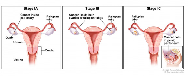
Cancer cells are found in the ovaries or fallopian tubes.
- Stage IA: Cancer is found inside a single ovary or fallopian tube. It is not found in the abdomen or on the ovarian or fallopian tube surface.
- Stage IB: Cancer is found inside both ovaries or fallopian tubes, but it is not found on the ovarian or fallopian tube surface or in the peritoneal fluid or washings.
- Stage IC: Cancer is found inside one or both ovaries or fallopian tubes and one of the following is true:
- Stage IC1: the capsule (outer covering) of the ovary ruptured (broke open) before or during surgery; or
- Stage IC2: the tumor wall ruptures prior to surgery or there is cancer on the ovarian or fallopian tube surface; or
- Stage IC3: cancer cells are found in the fluid of the peritoneal cavity (the body cavity that contains most of the organs in the abdomen) or in washings of the peritoneum (tissue lining the peritoneal cavity)
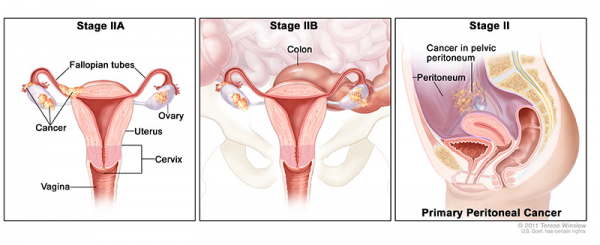
Cancer cells have spread from one or both ovaries or fallopian tubes and have spread to other tissues in the pelvis.
- Stage IIA: Cancer has spread from where it originated to other areas such as the uterus, fallopian tubes, and/or ovaries.
- Stage IIB: Cancer cells have spread to organs in the peritoneal cavity (the space that contains the abdominal organs)
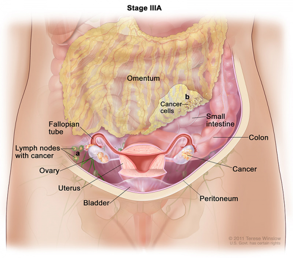
Cancer involves one or both ovaries or fallopian tubes. The cells have spread to tissues outside the pelvis or to the regional lymph nodes.
- Stage IIIA1: cancer has spread to the retroperitoneal lymph nodes (located at the back of the abdomen), but not to the peritoneal surfaces.
- Stage IIIA(i): The spread is 10 millimeters (mm) or smaller.
- Stage IIIA1(ii): The spread is larger than 10 mm.
- Stage IIIA2: cancer has spread microscopically from the pelvis to the abdomen and it may or may have not spread to the retroperitoneal lymph nodes.
- Stage IIIB: Cancer has spread to the peritoneum outside the pelvis and the cancer in the peritoneum is no more than 2 centimeters. Cancer may have reached lymph nodes behind the peritoneum.
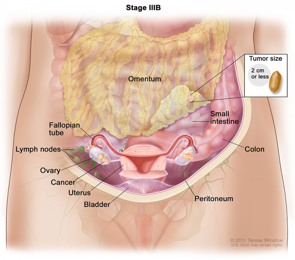
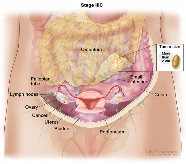
- Stage IIIC:Cancer has spread to the peritoneum outside the pelvis and the cancer in the peritoneum is 2 centimeters or larger. Cancer may have also spread to regional lymph nodes, the liver, or the spleen.
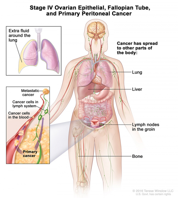
Cancer cells have spread to tissues outside the abdomen and pelvis. Cancer cells may be found inside the liver, in the lungs, or in other organs.
- Stage IVA: Cancer cells are found in extra fluid that builds up around the lungs.
- Stage IVB: Cancer has spread to organs and tissues outside the abdomen, including lymph nodes in the groin.
Oncologists also describe ovarian cancer by their grade. The grade describes the comparison between cancer cells and healthy cells when viewed under a microscope. This comparison of cells helps the doctor predict how quickly the cancer may spread. It can also help your health care team make better decisions about your treatment plan.
Ovarian cancer grades are as follows:
- Grade 1: The tissue is well differentiated. This means the cells look and are organized within the tumor like normal cells. In most cases, these tumors grow slowly.
- Grade 2: The tissue is moderately differentiated. It shares features between well and poorly differentiated. Grade 2 is not as commonly used as the other two grades.
- Grade 3: The tissue is poorly differentiated or undifferentiated. This means that all or most cells appear very abnormal and do not have any normal tissue structure. Oftentimes, these tumors grow fast and spread rapidly.